SOFTWARE
MorphoGraphX
MorphoGraphX is a software for image processing on surfaces. Time lapse data, typically from confocal imaging, is used to extract the surface shape of an organ of interest to create a curved image (2.5D) of the surface layer of cells. The method allows the time-lapse quantification of samples that are too difficult to segment in full 3D, but have too much curvature for a single 2D section. Development of MorphoGraphX continues in Norwich at the JIC, as well as in the Tsiantis group at the Max Planck Institute for Plant Breeding Research in Cologne, where Soeren Strauss is a primary developer.
MorphoDynamX
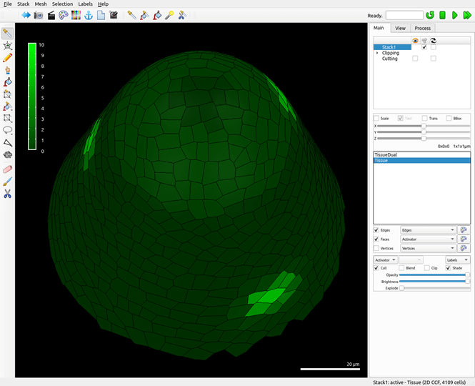
MorphoDynamX is a software for simulation modeling of development. It combines the image processing features from MorphoGraphX with a suite of tools for modeling cell division and growth, making it a unified framework for computational morphodynamics. It is based on the Cell Complex framework that provides a data stucture to represent geometry in any dimension, specialized for dynamic systems with dynamic structure. The Cell Complex framework is the thesis work of Brendan Lane, who is the project lead for MorphoDynamX.

A reaction-diffusion simulation of phyllotaxis on a growing shoot apex in MorphoDynamX
MorphoMechanX
MorphoMechanX is a software project led by Gabriella Mosca at the Centre for Plant Molecular Biology (ZMBP), University of Tübingen, Germany. The software is used for the finite element method (FEM) simulation of plant (and animal) tissue. It is implemented as an AddOn for the MorphoDynamX software, allowing the mixing of FEM and other MorphoDynamX models. Unlike many FEM frameworks targeted at biological tissue, MorphoMechanX is specialized for subdivision and growth.
An inflation simulation of pavement cells of Arabidopsis with the MorphoMechanX software (Add-On of MorphoDynamX)

MorphoRobotX
MorphoRobotX is a software for the control of micro-robotics actuators and force sensing equipment for experimental biomechnics. Like MorphoMechanX, is it implemented as an AddOn for MorphoDymanX.
Please visit the MorphoGraphX website for further information.
Costanza
Costanza is an open source segmentation tool developed to identify nuclei in 3D confocal data or cells with membrane marked (mainly used in 2D). It was originally developed in Java and is accessible via different interfaces:
ImageJ plugin that can be directly downloaded to the ImageJ plugin directory.
Jcostanza is a terminal-based java implementation for running batch jobs.
Pycostanza is a python implementation of the Costanza segmentation algorithm with extended features authored by Henrik Åhl.
Software authors: Henrik Åhl (formerly SLCU), Michael Green, Pawel Krupinski, Pontus Melke, Patrik Sahlin and Henrik Jönsson.
Organism-Tissue
The Organism-Tissue Simulator is a C++ software for simulating biological systems. It mainly focuses on systems with multiple cells/compartments, and includes biochemical as well as mechanical and cell proliferation rules for numerically simulating the system dynamics. The software is currently divided into two separate codes:
Organism used for simulating cells with geometries that can be described by a fixed number of variables and parameters. Examples are cells described by Spheres, Capped cylinders for rod-shaped cells, and Budding (yeast) shapes. It also can be used for simulations on geometries extracted from confocal data, where it uses information on cell volumes cell-cell connections (walls) and their areas.
Tissue used for simulating vertex-based cell geometries including finite element mechanical models. A cell wall (cell in 2.5D) is defined by a list of vertices defining a polygon.
Some of the features of the Organism-Tissue software are:
– It includes several numerical solvers for the ordinary differential equations, also with noise, and for stochastic simulations (Organism).
– It has a library of common biochemical, growth, division and mechanical rules.
– Models are defined within a specific model file – no need for recompilation when models are changed.
– The code is compartmentalised such that it should be easy for a programmer to define additional user-specific reaction- division- etc. rules.
– It is, and has been used to simulate growing plant tissue, and yeast and bacterial cell colonies.
– Includes visualisation and analysis tools.
– Includes an optimisation environment where data from multiple mutants can be used to define a cost function (only Organism).
The software is mainly developed at the Computational Biology & Biological Physics group at Lund University and the Computational Morphodynamics group at the Sainsbury Laboratory, University of Cambridge.
Developers (incomplete list): Henrik Jönsson, Patrik Sahlin, Pontus Melke, Pau Formosa-Jordan, Laura Brown, Pawel Krupinski, Behruz Bozorg, Niklas Korsbo, Ross Carter, Argyris Zardilis.
Tubulaton
This software is developed for simulating the dynamics and interaction of individual microtubule elements in 3D and confined within a closed surface (cell).
Software author: Vincent Mirabet, with contributions from Ross Carter, Tamsin Spelman, Pauline Durand and Cameron Gibson at the Computational Morphodynamics group at the Sainsbury Laboratory, University of Cambridge.
Biological Image Processing (BIP)
Biological Image Processing (BIP) is a general purpose software for the processing and analysis of biological images, with many operators particularly adapted to images of plant cells and tissues. The software is developed in C++ by the Modelling and Digital Imaging team led by Philippe Andrey at IJPB in Versailles. BIP is a command-line tool specifically designed for reproducibility in image analysis, facilitating the conception of complex pipelines and their application to large image datasets. The software transparently supports all image types (from 2D to 6D) and numeric formats (from 8-bit integers to 64-bit floats). The current version integrates >150 operators (image filtering, mathematical morphology, segmentation, quantification of objects and their contacts, etc.).
